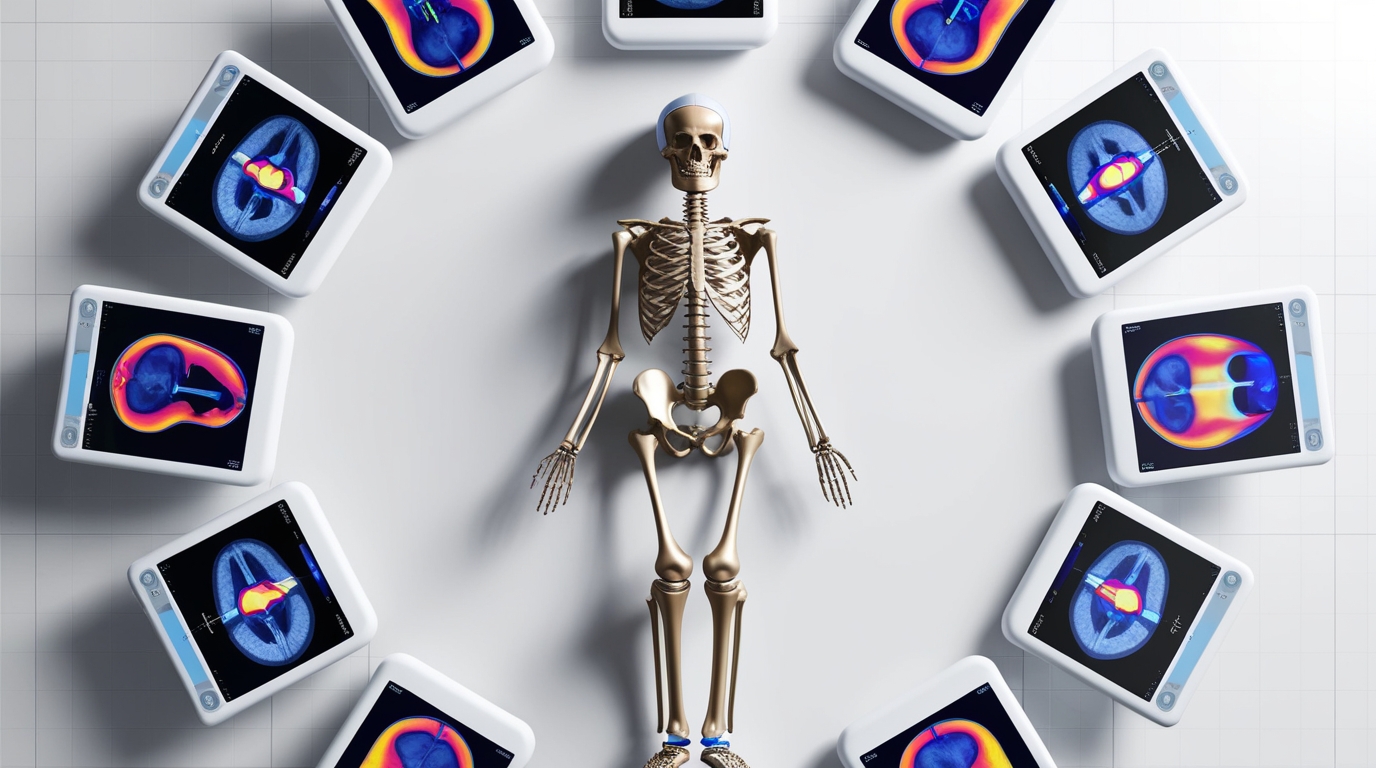
A CTscan or Computed Tomography scan is an advanced medical imaging technique. It combines x-ray measurements taken from different angles to create cross-sectional images of the body. It provides detailed views of internal structures, helping to diagnose a variety of conditions including cancer, cardiovascular diseases and neurological disorders.
The high-resolution images of CT scans improve the accuracy of surgical planning, biopsy guidance, and radiation therapy. Despite concerns about radiation exposure, the advantages of accuracy, diagnosis, and monitoring make CT scans an important tool in modern medicine, significantly improving patient outcomes and treatment efficacy.
Accurate diagnostics: CT scans provide high-resolution, cross-sectional images of the body. Enables accurate visualization of fractures, joint abnormalities and soft tissue conditions. The diagnostic accuracy of CT scans in detecting fractures is better than traditional X-rays. Especially for complex fractures of the hip, spine and shoulder girdle. According to a study published in the Journal of Orthopedic Trauma, CT scans improve fracture detection by 25% compared to plain radiography. This significantly reduces the risk of misdiagnosis and subsequent complications.
Preoperative Planning: Detailed anatomical information from CT scans is invaluable for preoperative planning. Surgeons can assess the exact location, size and orientation of fractures and the relationship between bone fragments. This information is important for developing accurate surgical plans and selecting appropriate fixation methods. For example, in spine surgery, CT scans help identify optimal placement of screws and other hardware, reduce the risk of nerve damage, and improve surgical outcomes.
3D Reconstruction and Virtual Surgery: One of the significant advances in CT technology is the ability to create three-dimensional (3D) reconstructions of anatomical structures. These 3D models provide a detailed view of complex fractures and defects. They help visualize them before surgery. Virtual surgical planning using 3D models allows surgeons to practice and refine their techniques. This will lead to more accurate and successful interventions.
Post-operative monitoring: They are essential for post-operative evaluation and monitoring. They allow for the healing of fractures and the diagnosis of any problems such as infections or non-union of bones. Accurate assessment of implant placement is critical in procedures such as total hip and knee arthroplasty.
Where incorrect positioning can lead to poor outcomes and the need for corrective surgery. A study conducted in the European Journal of Orthopedic Surgery and Trauma found that CT scans provide better assessment of implant alignment compared to conventional radiography.
Radiation Exposure and Safety Considerations: Although CT scans offer many advantages, it is important to consider the associated radiation exposure. Advances in CT technology have led to the development of low-dose CT protocols, significantly reducing radiation dose without compromising image quality.
For example, a comparative study in the American Journal of Roentgenology found that using modern low-dose CT techniques reduced radiation doses by 30-50%. Ensuring the judicious use of CT scans and adherence to radiation safety guidelines are essential to minimize risks, particularly in pediatric and adolescent patients.
CT scan has become an indispensable tool in orthopedics. Despite concerns about radiation exposure, advances in low-dose CT protocols have made this imaging modality safer for patients


